Blog, News, Advice and More!

Education Zone

Prevention and Treatment of Sweet Itch

Prevention and Treatment of Sweet Itch

Understanding Sweet Itch.  The weather is getting warmer, the sun is high in the sky, and your horse starts itching and rubbing his mane and tail. Sound familiar? Many owners dread the onset of summer because of the continuous threat of sweet itch. Sweet itch, or Summer Seasonal Recurrent Dermatitis, is an allergic reaction to the bites and saliva of Culicoides midges. It affects any breed or age of horse and usually occurs between the months of April and October when midges are most active. The threat of sweet itch should decrease during spring and autumn and disappear during the winter. The allergic reaction can range from mild to very severe, resulting in hair loss and bleeding scabs, which may lead to infection. Therefore, it is essential to recognise sweet itch as soon as possible, and take action to prevent it worsening or, if possible, prevent it from occurring at all. What are the symptoms of sweet itch? Sweet itch symptoms can vary greatly between horses, as some suffer more serious effects than others. Mild symptoms include itching and rubbing, usually around the mane and tail and along the top of the back. This can cause patchy hair loss on the coat and loss of mane and tail hair, which may lead to broken and damaged skin. If left untreated, the damaged skin can become sore and infected. In severe cases, sweet itch can extend under the belly, and down as far as the legs. There are some behavioural signs to look out for, too. Horses suffering with sweet itch may become easily agitated, restless or lethargic and show a lack of concentration during work. Vigorous swishing of the tail or headshaking is also common. How do I prevent sweet itch?Prevention is better than cure, and there are a number of measures you can take to minimise the risk of your horse developing sweet itch. Good management Midges are mostly active at dawn and dusk, so if possible, bring your horse inside at these times. Try protecting your horse in the stable by applying fly repellent or cover the stable door with fly-proof netting. Be aware that certain field conditions are more attractive to midges since they congregate near water or boggy marshes, so try not to turn out near these areas. Fly repellents It’s important to do as much as possible to keep flies and midges away. Using a fly repellent can do just that, and they are available as sprays, creams or gels, so there is something to suit everyone. Horses respond differently to each brand of repellent, so make sure you find the one that is right for your horse. Although usually very effective, repellents do wear off the skin over time, so they may need to be applied a couple of times a day, or every few hours if midges become particularly bad. Sheets and hoods Specially designed sweet itch sheets and hoods are now available which enable horses to be turned out without the need for frequent application of repellents. Special fabrics are often used that midges are unable to penetrate. They added bonus is that they also protect the hair, mane, and tail from being rubbed. How do I treat sweet itch? When sweet itch strikes, it is important to treat it as soon as possible. Keep irritated areas clean and dry. If the skin is sore or broken, apply a healing agent like calamine lotion or a nappy rash cream such as Sudocrem®  to help soothe irritation and soreness. Remember that these products won’t protect from further bites. Rubbing benzyl benzoate into the skin is also known to help (although this is not recommended for use on broken skin), or look for an alternative made from natural ingredients. With good management and care, sweet itch is a manageable condition, allowing you and your horse to enjoy the summer months. However, if the sweet itch becomes too severe, or you are worried about infection, it is always wise to consult your vet. For more information or advice on sweet itch, contact the Itchy Horse Company (Formerly known as the National Sweet Itch Centre), Tel: (01352) 840333; visit: HTTP://WWW.itchyhorse.CO.UK
An introduction to Cushing’s Disease

An introduction to Cushing’s Disease

Introduction to Cushing’s Disease (also known as PPID) What is Cushing’s Disease? Cushing’s Disease (also known as PPID, short for Pituitary Pars Intermediate Dysfunction), is a chronic progressive condition that shares some similarities with equine metabolic syndrome. It is one of most common problems affecting the hormonal (endocrine) system in horses and ponies and particularly in those over 15 years. Thanks to advancements in horse welfare, horses are living longer so Cushing’s Disease is becoming more prevalent. What causes Cushing’s Disease? Cushing’s Disease is caused by the overproduction of hormones by an enlarged and overactive pituitary gland ‐ a pea‐sized gland in the base of brain. The overproduction of ACTH (adrenocorticotropic hormone) by the enlarged Pars Intermedia (middle) part of the gland stimulates the adrenal glands to produce more cortisol (a steroid hormone). It is the increased levels of cortisol which causes most of the clinical signs associated with Cushing’s Disease. The condition progresses as the nerves connected to pituitary gland slowly degenerate What are the clinical signs of Cushing’s Disease? The most common signs of Cushing’s disease are recurring bouts of laminitis, which is the most serious complication, and hirsutism (excessive hair growth). Cushing’s Disease is the only condition known to cause hirsutism, which ranges from mild changes in coat shedding to the production of a long curly coat. Other symptoms include: excessive sweating; increased appetite; increased drinking and urination; pot‐bellied appearance; lethargy or more docile temperament; poor performance; lower immune function leading to recurrent infections eg skin, respiratory or dental problems; greater susceptibility to worms; loss of muscle condition, particularly over the topline; abnormal fat deposition, particularly around the Insulin resistance is also associated with Cushing’s Disease. How can I prevent or manage Cushing’s Disease? Cushing’s disease is a progressive condition so there is no known prevention. However, the risk of nerve damage may be lower in horses kept in good health with high levels of anti‐oxidants but this is unconfirmed. Similarly, early treatment following diagnosis may slow down the progression but, again, this is unproven. Given their lower immunity, horses with Cushing’s Disease should be on an effective worming, dental and vaccination regimen, plus any wounds should be frequently monitored for infection. Vitamin C supplementation (20 g per day) has been shown to increase antibody response to vaccines in aged horses, especially those with pituitary dysfunction or Cushing’s syndrome. See Feeding horses with EMS & Cushing’s Disease  How is Cushing’s Disease treated? There is no cure but effective treatment is available to address underlying hormonal imbalances. So far, Pergolide is the only licensed medication to demonstrate sufficient benefit and works by reducing the production of cortisol. Pergolide is available in a tablet form that can be hidden in feed. Associated conditions, such as laminitis, also need to be managed and treated accordingly. Horses with Cushing’s Disease may develop insulin resistance, so this will be managed in a similar way to EMS. Additionally, clipping will help to prevent excessive sweating if your horse grows a thick coat. What is the prognosis of Cushing’s Disease? There is no reason why a horse or pony with Cushing’s Disease, that is well managed, can live a long and normal life. Thank you to vet, Rachel Harrison-Osborne of Wendover Heights Veterinary Centre, for her input to this article. Helpful resources https://www.ed.ac.uk/polopoly_fs/1.18061!fileManager/dvepfactsheet‐cushingsdiseaseandequinemetabolicsyndrome.pdf https://www.laminitis.org/cushings.htm https://www.bhs.org.uk/welfare-and-care
Managing a Horse with Hock Arthritis – A Physiotherapist’s Perspective

Managing a Horse with Hock Arthritis – A Physiotherapist’s Perspective

If your horse has had a diagnosis of hock arthritis, there are many ways in which we can help the horse continue a working career and maintain a comfortable life. Adjustments may be needed in their husbandry and exercise programmes, but regular veterinary surgeon and complimentary therapist’s treatments will help to control any associated pain and discomfort. This article aims to give an overview on simple things you can implement into your daily routine to help with the management and associated problems with hock arthritis. As explained in the previous article The Horses Hock | Common Problems, Diagnosis and Treatment the hock is a very complex joint, which plays an important part in the performance horse. When problems arise in the hock joint the horse alters their gait to compensate, trying to avoid or minimise any pain or discomfort due to the arthritis. This results in compensatory hypertension and hypertrophy (overdevelopment) in the associated muscles, tendons and ligaments that are now having different stresses and strains applied to them. Due to a possible reduction in hock flexion, and also in an attempt to reduce concussive forces being applied directly through the hock joint, horses often compensate by circumducting (an outward swing) the affected limb during the swing phase of the stride cycle. This results in the muscles on both the medial (inside) and lateral (outside) aspects of the limb having different forces applied to them which can therefore become tight and overdeveloped. We also often see an increase in tension in the lumbar paravertebral region, gluteal region and hamstrings (see image below) – particularly the semimembranosus and semitendinosus muscles of the hind limb. Again, this is due to the horse trying to cushion and reduce the impact from the affected limb(s) striking the ground. The affects of which will often be seen in the ridden horse by being tense in the back, behind the saddle and having a shortened stride of the hind limb. There are many ways in which we can help support and maintain as near normal function as possible within these structures to help prevent further problems. Maintain Core Strength Horses will naturally try to limit and reduce the amount of pain or discomfort felt from an injury or joint disease by off-loading the affected limb. You may see them resting the affected limb more frequently. This off loading over time will have knock on effects on the core musculature which will affect the posture of the horse, and possibly lead to secondary issues. We can help maintain core strength and suppleness by performing baited stretches as part of our daily routine. Stand the horse square and slowly ask them to follow a treat around to the girth area either side, then the flank either side, outside the front fetlock either side, to the chest, between the front legs and then stretching up to the sky. It is always best to perform these exercises when the muscles are warm, so it is advised to do them after exercise, once tack is removed, but before the horse has cooled too much. If your horse is currently not in work then the muscles can be warmed by massaging the area prior to stretching. Click here for a more detailed guide on baited stretches. Massage Massage can be beneficial to help maintain suppleness within the muscles and prevent excessive tension, which can result in altered gait patterns. Regular application of massage, using effleurage – gentle stroking techniques, or petrissage a deeper kneading technique, paying particular attention to the lumbar paravertebral and gluteal muscle groups (muscles running along either side of the spine from behind the saddle region and over the rump) and the hamstring muscle groups (those running down the back of the hind limb and down to the hock). The Equilibrium Massage Mitt or Massage Mitt Hotspot can be particularly useful for massaging these areas. Manual massage can be tough on your hands if you are not conditioned to it, and these products can take away any worry of not knowing the exact techniques to use. The heat from the Massage Mitt Hotspot is very comforting for sore muscles and could also be applied to the hock joint itself before exercise to help warm the area and relieve initial stiffness. Massage can be performed regularly, if time permits, and if carried out after exercise as part of a routine it can help prevent tension building up. 20-30 minutes should be spent on either side of the horse 2-3 times a week (or more if time allows) to help maintain soft and supple muscles. If you find your horse is particularly uncomfortable when you are massaging a specific area it is always advisable to contact your veterinary surgeon or therapist for advice. For many of us, time on the yard is precious, especially through the winter months. The Equilibrium Massage Pad and the Massage Pad Heatsense provide an easy to use, effective massage (massage & heat from the Heatsense) for the muscles of the back and can be a useful tool to help aid the warm-up process before you ride. One of the biggest benefits of these products is that you can carry on other yard chores whilst your horse has their massage. Poles The use of pole work to increase range of motion within the hind limb can be very useful, but needs to be performed with care, your horse’s ability to perform the exercise correctly and being able to move over the poles without knocking them should be taken into consideration when deciding to use poles as part of the rehabilitation. Your Veterinary Surgeon or therapist will be able to advise you more if pole exercises are suitable for your horse. Something as simple as placing a couple of poles at intervals on your walk to and from the field will help to encourage your horse to use the limb through a greater range of motion. When using poles, it is important to take into consideration your horse’s fitness and ability – keep it simple. A single pole walked over cleanly can be much more beneficial than a grid of poles which your horse struggles to perform. Start simple and increase the number of poles over time. Try to always have someone on the floor watching your horse’s movement over the poles, we aim to have a regular stride length and equal limb flexion / extension through the pole configuration and for any horse diagnosed with hock arthritis I prefer to work with the poles in straight lines, rather than on a curve so we keep limb loading as equal as possible. Changes to Management When manging a horse suffering from hock arthritis alterations to their husbandry and exercise regimes may be required to help achieve the best results, with regular assessments from your veterinary surgeon, therapist and farrier the best outcomes can be achieved. You should avoid long periods of restricted mobility and try and keep exercise levels regular throughout the week, avoiding overworking on hard or deep surfaces and be conscious of ground conditions and the terrain you’re working your horse on – harder surfaces and hills will increase the forces applied through the affected joints. As with any Equine injury, ailment or disease if you are unsure at any point about what is appropriate for your horse we advise you refer back to your Veterinary Surgeon or Musculoskeletal Therapist for advice. Thank you to Catherine for writing this article: Catherine MarshallVeterinary PhysiotherapistAdvCert VPhys DipAVN(surgical)RVN Cert Clin Ed Acc Midx MRAMP, MIRVAP
An introduction to Sarcoids

An introduction to Sarcoids

What are sarcoids? Sarcoids are benign skin tumours that can be found in horses, donkeys and mules, they are generally not life threatening but do destruct the skin cells that are surrounding them. They appear in different forms on a horse and are most commonly found on the abdomen, inside the backs of legs, the chest and around the ears and eyes. Normally at the sight of previous traumas and where flies congregate. Even though there are 6 different types of sarcoids they all start small and grow at different rates, when they enlarge the skin may ulcerate that can attract flies and cause an open sore. Why does a horse get sarcoids? Sarcoids are caused by an infection the virus Bovine Papilloma Virus (BPV), it is a virus that originates from cattle but is spread by flies. All horses may be infected by BPV in their life, however not all of them will be susceptible to the virus and present with sarcoid growth. How do I know if my horse has sarcoids? Not all lesions that appear on the skin are sarcoids so a vet should be able to diagnosis whether it is a sarcoid or any other skin problem.  If the vet is unsure they can take a biopsy to confirm the diagnosis, this is the last resort diagnosis as a biopsy is invasive. As to know if your horse has the gene that makes them susceptible to sarcoids there is a genetic test that can be carried out however these are not used very often by vets as they can be inconclusive and are expensive to run. How can I prevent sarcoids? There is no easy prevention of sarcoids as there has not yet been a vaccine developed for the disease. Making sure your horse’s immune system is at its strongest in order to help fight the virus and boost the immune system. How are sarcoids treated? There are many ways that sarcoids can be treated and as they all react differently to treatment. The main way of treatment is the use of Liverpool cream, a chemotherapy cream created at the University of Liverpool. This takes place over a number of weeks and can become expensive as the  cream can only be applied by a vet.  The cream is applied every day for a week or every other day for 2 weeks although this may change on a case by case basis.  It is method that makes the sarcoid look worse before becoming better, as it kills all the cells from the area that it is applied to and after a long period of time the sarcoid falls off along with all of the skin cells containing the virus. This then leaves a raw patch of skin exposed (see picture on right), which is why the best time for Liverpool treatment is in the winter where no flies can infect the exposed patch of skin.  There  are many success stories of it fully getting rid of sarcoids although others say that it returned, again reiterating that every sarcoid is different and may have a different reaction to treatment. Another way is rubber banding this is specific to certain types of sarcoids as it is not possible to get a band around every one as some lie flat. But if a band can be placed around it, then the circulation to the sarcoid will slowly be cut off until it dies and falls off.  Along the same lines as this,  there is also laser surgery and Cryosurgery.  They all aim to remove the sarcoid cleanly leaving no trace. Laser is the least invasive using lasers to remove it, Cryosurgery uses a repeated rapid freezing method which is time consuming. Alternatively there are homeopathy treatments that have worked in some cases but also have limited success as they tend to need harder treatments to remove the sarcoids. However there has been proven success with Bloodroot treatment which is a plant extract, used in a similar way to Liverpool cream, the picture on the left shows a pony who had bloodroot treatment and the results after 3 weeks (2 weeks of treatment with 1 week off in between), for this pony however, the treatment did not completely get rid of it. Do I need to treat my horse’s sarcoids? Not necessarily if the lesion is not getting bigger or is in a place that will not affect the horse then it is up to you if you leave it. However when the sarcoid is smaller it is easier and quicker to treat so early treatment is recommended. Discuss with your vet which is the best method of removing for your horse and its type of sarcoid.
Colic in horses - causes and prevention

Colic in horses - causes and prevention

What causes colic? Colic can be caused by gut “spasms” (e.g. due to a change in diet), impaction of partially-digested food material or sand, stretching of the gut wall by gas, twisting of the gut so that its blood supply is cut off, displacement of a segment of gut from its normal position, stomach ulcers and fatty tumours wrapping around the gut and strangulating it. Horses are prone to colic because of their unusual GI tract anatomy (particularly the length) and the way their gut works.  It is important to note that many other conditions, e.g. urinary problems, azoturia etc, can present as colic. What are the clinical signs of colic? Mild colic Moderate colic Severe colic ·         Lip curling   ·         Flank watching ·         Restlessness ·         Pawing the ground ·         Dullness ·         Frequently posturing to urinate   ·         Lying down and getting up ·         Lying on their side for long periods ·         Violent rolling   ·         Sweating ·         Rapid breathing How can I prevent colic? Some causes of colic are preventable if good stable management and a regular daily routine are followed: Use an effective worming programme to control parasite levels. Ensure your horse has a constant supply of fresh and drinkable water. Feed small quantities and use good quality, high fibre, feed stuff and include plenty of forage in your horse’s diet. Be consistent with your feeding routine and make gradual changes to your horse’s diet. Ensure your horse has a consistent turn-out and exercise regime. Turn out on good quality pasture, avoiding poor or overgrazed fields or, conversely, lush spring grass. Have an effective dental programme in place because tooth problems can cause digestive issues if food isn’t chewed properly. In sandy areas, avoid feeding horses from the ground as they may ingest large amount of sand. Reduce stress levels by ensuring your horse has a consistent routine and environment and be mindful of stress when travelling your horse. If your horse has a habit of eating his bedding, ensure he is bedded down on something other than straw; this is particularly important if your horse has to suddenly undergo a prolonged period of box rest. Habits such as crib-biting and windsucking will increase the risk of colic and some horses are more prone to recurrent episodes of colic Thank you to vets, Rick Farr and Nikki Pursey, of Farr & Pursey Equine Veterinary Services, for their input to this article. Helpful resources The Royal School of Veterinary Studies – colic information sheet The University of Liverpool Equine Hospital
Common Equine Eye Complaints

Common Equine Eye Complaints

The equine eye The eyes of the horse are located laterally (either side) of the skull which maximizes the horse’s field of vision. The equine eye is a very delicate structure and in comparison to other species, does not tolerate injury or disease at all well. For this reason it is highly recommended that veterinary advice is always sought as soon as possible in any problem relating to the equine eye. First of all a complete history of your horse will be taken, his age, breed and any previous illnesses noted. Current management of the horse is then determined followed by the details of the current problem. The eye is a complex organ and there are numerous structures inside the eye which the vet will examine. First of all, the gross appearance of the eyes are assessed, checking for symmetry, swellings, overflow of tears, etc. His vision is then assessed by the menace reflex (does the horse blink if an object moves towards his eye?) and the pupillary reflex (a light is shone in the eye and the pupil should constrict). Assessment of the Equine Eye In a darkened stable, an opthalmoscope enables the vet to visualise the interior of the eye. An assessment of the following structures is performed: Cornea Anterior chamber Lens Iris Vitreous body Retina Optic disc A normal, healthy eye will be open, clear and bright with no discharge. The conjuntivae will be pink and there will be no swelling present. A painful eye will present with some or all of the following signs: Blepharospasm (eye clamped shut) Lacrimation (tear overflow) Chemosis (swollen conjunctivae) Discharge (yellow, white or green) Photosensitivity (sensitivity to light) Corneal oedema (cloudy eye) Miosis (Constriction of the pupil) If your horse presents any of the above clinical signs you should phone your vet immediately. Some Common Conditions Corneal Ulcers This is a very common condition seen in all type of horses or ponies and is extremely painful. Your horse’s eye maybe swollen, clamped shut, watering profusely and may have a cloudy appearance. Due to the pain he is experiencing, your horse will not allow you to open his eye manually and may even become naughty if you persist in trying. A corneal ulcer is where the surface of the eye is damaged, usually caused by some kind of trauma or foreign body. Your vet will stain the eye to check for damage. An intact, undamaged cornea will not take up the stain whereas most corneal ulcers will stain green quite clearly. Treatment consists of frequent application of serum, antibiotics and pain relief direct to the eye. Oral pain relief medication may also be administered if required. Turn out is permitted only with a fly mask to prevent damage to the back of the eye from the sun and secondary infection from flies or dirt. Some corneal ulcers can be very persistent so prompt veterinary attention is essential. Equine Recurrent Uveitis Also known as “moon blindness”, this condition is an important condition in that it can recur throughout the horse’s life causing significant painful episodes which, in extreme circumstances, can lead to the removal of the eye. This condition is often mistaken for a traumatic injury due to the amount of pain the horse is in and the swelling around the eye. ERU’s primary cause can be a virus, systemic disease or trauma. It is thought to be an immune-mediated disease where the structures inside the eye are inflamed. Often debris can be seen in the front of the eye, the cornea may be cloudy and the pupil constricted. Examination with the opthalmoscope is often resented due to increased sensitivity to light and the eye does not take up the stain. Treatment consists of pain relief, topical steroids, antibiotics, serum and atropine (to dilate the pupil). This condition can be frustrating due to random repeat episodes causing significant pain and requiring aggressive treatment. It is thought that sunlight and wind can aggravate an episode so turn out is permitted only with adequate eye protection such as a fly mask with an eye patch sewn in. Conjunctivitis Also a common but not so painful a condition, conjunctivitis is a bacterial infection of the eye. As an owner you will find your horse has an ocular discharge which is usually yellow or green in colour. The conjunctivae will be inflamed and there may be some swelling but in general the horse is not too worried. Your vet will stain the eye to check for corneal damage and usually just dispense antibiotic eye drops. It is recommended that sterile water / saline soaked cotton wool swabs are used to clean the discharge away twice daily. In the summer months, ocular discharge will attract flies which exacerbate the condition, therefore it is recommended the horse is turned out at night, or otherwise in a fly mask. Blocked Tear Ducts This condition tends to worry the owner more than the horse. It presents as a persistent, watery, ocular discharge which despite regular cleaning does not settle down. It usually occurs in the warmer months and is very obvious when the horse is brought in from the field. There is no pain or swelling with this condition unless a secondary bacterial infection has occurred. There can be hair loss in the area where the watery discharge lies or just matting of the hair in this area. Normally tears produced by the eye, flow down the nasolacrimal duct and drains into the nose. However the tear ducts are very fragile structures which are lined with a soft membrane. If damaged this lining can stick to itself and block the duct resulting in an overflow of tears from the eye. Although not a serious condition, the ocular discharge does attract flies which in turn can cause a bacterial infection. Two treatment options exist, the first is management rather than treatment. The eyes can be wiped cleaned twice daily with sterile, soaked swabs and a fly mask used whenever the horse is turned out. Antibiotic eye drops maybe dispensed. The second option requires the horse to be sedated so that the tear ducts can be flushed with sterile saline. This is not a painful procedure but the horse does feel some discomfort. Steroid / antibiotic eye drops are then dispensed to reduce any inflammation within the ducts. Many horse owners feel happy managing this condition and tend to keep the flushing procedure as a back up plan. Flushing the tear ducts is obviously more costly and can be disappointing in that there may be a recurrence in the ducts becoming blocked. Fly Mask Selection For many eye complaints, treatment includes protection of the eye from sunlight and insects. For ERU ('moon-blindness') a mask with an eye patch for 100% protection is normally recommended. For most other conditions, a good quality fly mask is recommended by many vets to their clients. It should be looked at that the fly mask stays well away from the eye, is comfortable for the horse and offers UV protection too. Summary There are several common eye conditions, which can be very painful for the horse. Horses' eyes are unusually delicate, and this, combined with the horse's reliance on the sense of sight, means that all symptoms of a potential eye problem should be treated seriously. It is strongly recommended that veterinary advice is always sought as soon as possible, to assess the condition and start treatment. By Claire McKinstry MRCvs  
Coping with a cold back

Coping with a cold back

Christine East, a McTimoney Animal Practitioner, shares her expertise on how to care for your cold-backed horse. An increasing number of horses are being diagnosed as ‘cold-backed’, and the problem is much more common than many horse owners think. So what does it actually mean, and how can it be treated? What does ‘cold-backed’ mean? The term ‘cold-backed’ is used to describe a horse displaying symptoms of a sensitive or painful back. These symptoms can range from very mild, such as discomfort when the girth is tightened, to more serious, lasting until the horse has warmed up and the muscles are relaxed. McTimoney Animal Practitioner, Christine East has treated many cold-backed horses. “There are lots of different interpretations of the term ‘cold-backed’, and it can be used to cover a range of different symptoms,” she says. “However, it is important to remember that every horse is an individual, so they may display different signs to other horses with the same problem.” What to look out for Christine stresses that it is common for horse owners to worry about confusing a cold back with their horse’s behaviour towards activities, such as tacking up and grooming. However, there are definite signs to look out for, which include: Soreness or sensitivity to grooming over the backReacting to the saddle, such as dipping or bridging the back when it is put onDiscomfort when the girth is tightenedRefusal or reluctance to let you mount from the groundStiffness through the back during the first few minutes of work What is the cause of a cold back? Christine suggests that one of the underlying problems is evolutionary. “Horses didn’t evolve to carry people on their backs and compensate for the extra weight. This means they can develop sensitive nerve endings or a misalignment in the spine as a result. Some of the most common causes of a cold back include: pressure from a poorly fitting saddle; aggravation of previous injuries to back muscle; or problems with your horse’s teeth or feet, which cause them to readjust their posture. Another cause, which I think is often overlooked, is the posture and position of the rider,” says Christine. “I often have calls from owners whose horses all seem to have developed the same back problem and it is caused by the horse having to compensate for the rider’s bad posture. When having your horse treated, it is sometimes worth seeing a back specialist yourself!" Treating a cold back As there are such a range of symptoms for a cold back, it is important to consult your vet as a first port of call, who can rule out any injury or more serious problems. They can then refer your horse to a physiotherapist, chiropractor, or McTimoney practitioner, as necessary. “Symptoms associated with a cold back are not usually too difficult to treat,” says Christine. “Most treatments work to relieve spasms or misalignments in the spine, which cause a pull in the surrounding muscle tissue and result in soreness.” When treating a cold back, it is also advisable to have your horse’s feet and teeth checked, to ensure that there aren’t any problems here that are causing knock-on effects. Prevention is key There are certain preventative measures that can be taken to help stop your horse developing back problems in the future. Have your saddle checked annually, because a horse’s shape can change throughout the year and may not be the same as when the saddle was fitted. Always warm up properly before exercise and remember to cool down when you have finished – this allows your horse’s muscles to adjust to your weight and avoid any damage. If your horse is particularly sensitive, tack up and walk him around for five minutes before mounting – this will give the muscles a head start and ensure he is comfortable when you are mounting. In addition, always use a mounting block rather than mounting from the ground, as this can place extra stress on the back. Often, when a horse won’t stand still for mounting, it is an indication that he finds it uncomfortable, so take care as you get on. When schooling, ensure you do sufficient work on both reins – this will benefit the whole body, not just the back, and will help your horse to remain flexible through both sides of his body. A cold back is treatable “The good thing to remember is that the problems associated with a cold back are almost always treatable,” says Christine. As with everything though, prevention is better than cure, so taking good care of your horse’s back is important, and will help him stay fit and healthy and able to perform at his best.
Do horses need extra electrolytes?

Do horses need extra electrolytes?

What are electrolytes? Present in small amounts, electrolytes are positively charged ions that are dissolved in the blood and the fluid that exists between cells and sweat. The principal electrolytes are sodium, potassium, chloride, calcium and magnesium. How do horses get their electrolytes? Most horses receive most of their daily electrolytes from forage.  Calcium and phosphorus are also commonly added to compound feeds and mixes.  However salt can be low in forages and are not added in high levels into compounds.  Hence extra salt is routinely offered as licks or in the feed. Horses on low forage diets, such as those receiving a lot of hard feed, or those on restricted diets due to being good-doers, will have a lower electrolyte supply than horses with good fibre levels in their diet. Many working horses receive added electrolytes as a supplemented to their diet. Why are electrolytes important? Electrolytes are responsible for the correct function of nerves and muscles.  They also help maintain normal hydration and are integral in maintaining the functioning of the digestive system, and the other vital organs. Sub-optimal electrolyte status can lead to muscle problems and poor performance.  Since electrolytes cannot be stored in the body, the amount available to the horse is dependent on electrolyte intake compared to natural losses through sweat, urine and faeces. Electrolytes and sweating Sweating removes heat generated by muscles during exercise.  A fit horse efficiently removes heat from where it is not wanted in the muscles, out to the skin where it is lost through sweat- where the process of evaporation from the skin cools the horse. Equine sweat is hypertonic – that it is contains more salts than internal body fluids – which means that a sweating horse loses electrolytes as well as water.  Typically a working horse can lose 7-8 litres in an exercise bout. The amount of sweat produced by an individual depends on temperature, workload, temperament and stage of fitness. One litre of horse sweat contains around 3.5g of sodium, 6g of chloride, 1.2g of potassium and 0.1g of calcium I litre of sweat is estimated to dissipate heat from 1 –2 minutes of extreme work or about 5 minutes of sub-maximal work, summer or winter  When do horses need extra electrolytes? Whenever a horse works hard, summer or winter. Electrolyte use in the days before a competition will stimulate water intake such that the horse enters the competition fully hydrated. (long term mega-dosing is wasteful as any excesses cannot be stored) Before during or after travelling: offering water or an isotonic solution every couple of hours on long journeys should help avoid unnecessary fluid* and/or electrolyte deficits before the competition has begun. Before and during moderate to intense work: Research has shown that correct administration of good quality electrolytes can increase the time it take for a horse to fatigue by nearly 23%. (On the day of competition or exercise bout, the final offer of electrolytes should be made together with water, at least 4 hours before exercise.) Horses on low or restricted forage diets (i.e. less than 30% of total diet as forage) will receive a diet inherently low in electrolytes.. Equally a new forage source could be markedly different to the previous source, and cause problems.
Electrolytes and Gastric Ulcers

Electrolytes and Gastric Ulcers

Salt, electrolytes and gastric ulcers The use of electrolytes in relation to gastric ulcers has been mooted since a study in endurance horses showed those receiving a concentrated electrolyte paste once per hour for eight hours showed higher rate of gastric lesions than those receiving a placebo.  Extreme use such as this is not routinely recommended.   It is necessary to replenish lost salt and electrolytes after sweating, because sweat contains both water and electrolytes; sweating cools the horse by the process of the evaporation of water from the skin, but in losing water the horse also loses electrolytes, which must then be replenished through the diet. Ensuring a sound base of forage in the diet is essential – forages themselves are a source of electrolytes, and they also create a reservoir of water bound with the fibre in the hindgut that acts as a fluid reservoir when sweating. Supplementary electrolyte requirements are highly individual, dependent on the duration and intensity of the workload, the core ration as well as the environmental conditions.  Electrolyte tips: Maximise forage intakes where possible.  If travelling or on restricted forage diets consider offering forage in a  way that slows consumption such as double walled hay nets or the use of a forage block. Consider providing salt blocks at home; Always follow the supplement manufacturer’s recommendations for use Always offer fresh water when administering electrolytes Never administer electrolytes on their own into an empty stomach – try mixing them with some type of fibre – e.g. a small amount of alfalfa
Essential Hoof Care

Essential Hoof Care

The structures in a horse’s feet are responsible for supporting the full weight of the horse over a small area. Routine foot care is therefore extremely important, as any problems in the feet can be extremely detrimental to mobility and health. Structure of the foot Coronary band This is located at the top of the hoof and is responsible for creating horn that makes up the hoof wall. Periople This is the outer layer of the hoof that forms a protective covering on the hoof wall. It is responsible for regulating moisture content in the horn, secreted from the perioplic ring above the coronet. Hoof wall The hoof wall is the exterior of the hoof, made from a keratin-based substance.It provides a hard protective layer around the internal parts in the foot. It takes 9-12 months for the hoof to grow from the coronary band to the toe. In order for the horn to grow correctly and form a healthy foot, the horse must be provided with a good diet and be in good health. These factors must be checked if the horn starts to become brittle and weak or if the foot looks badly formed. A feed supplement of biotin may be helpful to promote good horn growth. Sole This is a tough structure that provides external protection to the sensitive sole underneath. It is slightly concave and is not weight bearing. Frog The frog has an extruding triangular structure which extends from the heel to halfway down the foot. Its function is to absorb concussion, provide grip and be a weight-bearing surface for the foot. It also ensures that a healthy blood supply reaches the foot. The grooves along each side of the frog allow for expansion when it makes contact with the ground. Inside the foot Sensitive sole This is found underneath the pedal bone, within the insensitive sole. It produces the new cells that replace lost layers of the insensitive sole. Digital cushion The digital cushion is found between the pedal bone and deep flexor tendon. It is an elastic, fibrous pad that absorbs concussion from ground impact. It also helps to push blood back up the leg. Lateral cartilages These are attached to the pedal bone and serve to protect the coffin joint. They also help absorb concussion. Laminae The insensitive laminae are supportive structures that attach to the hoof wall and interlock with the sensitive laminae. The sensitive laminae then attach and support the pedal bone. The divide between sensitive and insensitive laminae can been seen as a white line on the sole of the foot. Conformation This is extremely important, as the feet are obviously essential to the horse! They should be even and round in shape and in proportion with the rest of the horse. The fronts should be of equal size and shape and so should the hinds. The front feet should slope forwards and be at a 45 degree angle to the ground, and on through the fetlock and pastern. The hind feet should be at an angle of 50-55 degrees to the ground. The hoof wall should be smooth and free from cracks. Any lines could indicate poor nutrition or past cases of laminitis. Poor conformation in the feet can result in strains to tendons and ligaments, tripping and bruising. Many foot conformational faults can be improved by a good farrier and over a period of time. Routine care Apply hoof oil every other day during the summer to help prevent splits and cracks Pick out feet every day with a hoof pick Check shoes for wear and tear and signs that a farrier is needed – such as risen clenches, pinching across the bulbs of the heel, overgrown and misshapen feet Check unshod horses for splits, cracks, flares and overgrown misshapen hooves Ensure that the farrier attends shod feet every four to six weeks, and unshod feet every six to ten weeks Farrier Great care must be taken when selecting a farrier, so ask your vet for recommendations. Correct trimming and shoeing is vital to the horse’s welfare, and any mistakes can lead to serious, lasting damage. The horse’s feet should be correctly balanced whether shod or unshod. Balance is important as inaccuracy can lead to lameness and aggravate navicular syndrome and laminitis. It can affect the whole movement and development of the horse and cause ongoing problems. Shoes are not always needed. It depends on the amount and type of work the horse is doing. Sometimes only front shoes may be needed. The farrier will be able to advise on the best option for the individual horse. Checklist Always check the horse’s feet after the farrier has visited The horse should be sound. If he is even slightly lame or lame a few days later, the farrier must be recalled immediately to address the problem. Check the balance of the feet. The angle should be around 45-50 degrees from the ground at the front and 50-55 degrees from the ground at the back. The angle through the pastern, fetlock and hoof should be 45 degrees. When viewed from the heel, with the foot raised, the sides of the foot should be level. With the foot on the ground both sides of the hoof should be of equal length. If shod, the shoe should fit the foot with no gaps between the shoe and the foot. The clenches should be about one third of the way up the hoof wall from the floor. They should be in a straight line and be flush with the hoof. The toe clips should also be flush with the hoof wall. The sole of the foot should not be touching the ground in unshod horses The sides of the frog should be trimmed. The frog should be level or slightly below the edge of the hoof wall. If any deviations from the checklist are found, speak to your farrier. There may be a reason for this, such as correction of conformational defects, but the feet may need to be re-checked. Common ailments in the feet In most cases of lameness, the cause is usually found in the foot. Bruised soles These are caused by an injury to the sole of the foot, usually by standing on a hard object or concussion from hard ground. They can also be due to poor trimming or shoeing. Symptoms are acute lameness that gets progressively worse, red or bruised areas seen on the sole, and reaction to pressure on the sole due to pain. The treatment is to restrict movement and keep on a soft surface – a deep bed in a stable, sand school or woodchip area until sound. If in severe pain, call the vet who may prescribe anti-inflammatory drugs and check for any infection. Thrush This is caused by continuous exposure to a damp environment without sufficient care and attention to the feet, such as poor stable management, wet or damp bedding, and wet, muddy fields. It is a bacterial infection and if left untreated, it can move to the sensitive, internal structures in the foot. Symptoms are a black, smelly discharge around the frog, and possibly lameness if severe. The treatment is to scrub out the foot and apply eucalyptus oil (available from most chemists) repeatedly along the grooves of the frog until it clears. The farrier should trim the sides of the frog to remove any damaged tissue. If there is infection and lameness call the vet and follow the advice – it may need poulticing. To prevent thrush, keep the feet clean, scrub them out and apply eucalyptus oil at least once a week during the winter, and when necessary in the summer. Make sure that there is a dry area in the field, for example hard standing, if the horse is out all the time. Make sure bedding is kept clean and dry. Seedy toe This is the separation at the white line. It usually starts at the toe and gradually progresses up the hoof wall. The hole becomes filled with white, dead material. It normally occurs when the toes are allowed to become too long, but it can be a result of laminitis or of concussion on hard ground. This condition needs to be managed by regular, correct trimming by a farrier – the hole will then grow out. Some of the tissue may need to be cut away and packed with putty. There may be an infection so, if the horse is lame, call the vet, as antibiotics may be needed. The foot may then need to be tubbed with water and Epsom salts and poulticed. Laminitis This is caused by several factors, but the main reason is an overload of soluble carbohydrates in the digestive system (see the All About Pets leaflet, Laminitis (H14)). Symptoms are a reluctance to move, increased digital sesamoid pulse, walking heel to toe, and leaning back onto the hind feet. Call the vet immediately and follow the treatment plan given. Remove the horse from grass and take him into a deep bed of shavings, cardboard or sand until sound. To prevent laminitis, a properly formulated high fibre diet is necessary with strict weight control, and regular farrier attention. Infections in the foot (pus in the foot) This is caused by puncture wounds, seedy toe, or bruising. It is the most common reason for lameness. Symptoms are an extreme lameness due to the inflammation in the foot, increasing pressure against the hoof wall, causing pain. There is an increased digital sesamoid pulse in the affected hoof, and reaction to pressure on the infected site due to pain. Call the vet, as the infection (pus) should be released from the foot by digging out the infected area. This can also be done by a farrier. The horse will be sound or at least almost sound after this procedure. The foot will then need to be tubbed and poulticed to draw out the rest of the infection. If not treated, the leg can begin to swell and the infection can spread through the foot and burst out of the coronary band. In extreme cases the vet may prescribe antibiotics alongside practical procedures. Also, ensure that the horse is covered for tetanus, as puncture wounds are an ideal way for tetanus to enter the body. A vet should see all puncture wounds to the foot because if they are deep enough, they can infect the pedal or navicular bone. This is a serious condition and needs surgical attention. It can cause damage to sensitive, internal structures including tendons and could cause permanent lameness. Nail bind/prick This is caused by the farrier putting a nail too close to the sensitive part of the foot (nail bind) or actually piercing the sensitive part of the foot (nail prick). Symptoms are lameness after shoeing, either immediately or up to a couple of days later. To treat it, the farrier needs to remove the nail and the foot should be tubbed and poulticed as with a foot infection. Call the vet if lameness continues, or if the farrier recommends it. Check tetanus vaccinations are up to date. Sand/grass cracks A sand crack starts at the coronet band and works down, whereas a grass crack runs from the ground towards the coronet band. Both are caused by poor foot conformation or condition, poor or irregular farrier attention, or an injury. Call the farrier for treatment. The cracks can be stopped from spreading by marking a groove in the hoof wall above or below the crack, or by putting clips around the start of a grass crack. With regular, correct farriery, the cracks should grow out. To prevent cracks, ensure regular, correct farriery. A dietary supplement of biotin can also promote good hoof condition and growth. Original article produced by The Blue Cross Organisation. Read more at www.bluecross.org
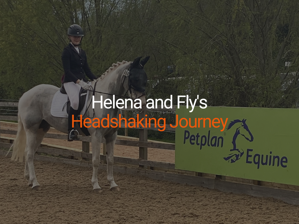
Headshaking - Common Myths Exposed

Headshaking - Common Myths Exposed

By Dr Katy Taylor,De Montfort University Head Shaking is not a single condition One of the most frustrating things about headshaking is that each case is unique. So, what causes headshaking in one horse may not be the cause in another. It’s important to recognise that headshaking is the symptom of a problem with the horse rather than being the problem itself, although it is often treated as such. There are over 60 known causes of headshaking, including ear, eye and respiratory problems, which makes a veterinary visit all the more important. Simply assuming your horse is an untreatable head shaker may cause more harm. Likewise, be wary of products that claim to cure all head shakers. Headshaking is not a behavioural or management problem Because headshaking often only arises when the horse is ridden, it is commonly attributed to tack problems, bad riding, or resistance. However, studies in the US and UK have shown that around 50% of head shakers also show symptoms at rest. So, whilst exercise always worsens the symptoms, most owners find that changes in tack or schooling have little effect. Researchers at De Montfort University and the University of Lincoln investigated head shakers and non‑head shakers and found no important differences in their diet, use or turnout. The relative unpredictability of the condition and symptoms suggest that the headshaking is not a behavioural one unlike weaving or nodding. Not all headshaking is caused by sunlight (photic headshaking) Head shaking has often been attributed to a reaction to sunlight, but whilst this may be so for some, especially animals in more sunny places like California, many European head shakers seem relatively unaffected by light. Riding indoors or at dusk can still bestow benefits other than by restricting exposure to light so the best test for the photic form of this problem is to carefully cover the horse’s face whilst exercising. Headshaking is not horse hay fever The seasonal nature of headshaking (50% of head shakers only have symptoms from March to October), together with the signs of nasal irritation, has led many to believe that headshaking is a respiratory disorder similar to human hay fever. However, vets remain sceptical because, in general, hay fever medication is not very effective. In addition, vets rarely find clinical signs of an allergic reaction. Although headshaking may be in response to particles in and around the nose, an allergic reaction may not necessarily be occurring. So what does cause headshaking? Given there are a variety of causes, a large proportion of head shakers are thought to be suffering from some kind of pain originating within the head. Nerve pain (neuralgia) has long been considered, ie. damage to the sensory nerves of the head, which may have resulted from physical damage to the nerves or following infection from certain viruses. The involuntary headshaking movement could be a response to the misfiring of a faulty nerve and the rubbing of the face in an attempt to relieve the discomfort caused. Facial neuralgia could account for the general hypersensitivity around the muzzle that many head shakers experience, particularly to rain, snow and wind. In some cases, exercise or particles may be causing the blood vessels in the nose to swell and ‘squash’ the nerve, causing it to misfire. The reasons why some horses head shake and others don’t are not yet known. Frequently the onset of their problem has been traced back to the beginning of the horse’s working life so the many changes that occur within the horse or in its environment during this time may hold part of the answer. Because the problem is often not apparent until the horse is exercised, headshaking could have arisen earlier when the horse was very young but has lain undetected until the horse was backed. Future research into how we can prevent headshaking in foals may be fortuitous once we know more about the causes of headshaking.
Kissing Spine in Horses

Kissing Spine in Horses

Over recent years ‘Kissing Spine’ seems to have become a more widespread and commonly diagnosed condition. The good news is that with the correct treatment and/or surgery many horses are making near to full recoveries and it doesn’t necessarily mean an end to their ridden career whether this is as a happy hacker or a competition horse.  What is Kissing Spine? Kissing spine is a disease of the spine whereby the spaces between the upper dorsal processes of the vertebrate become reduced and the vertebrate actually touch, or ‘kiss’, causing bone to bone contact and in some cases damage to the ligaments within the spine which causes the horse great discomfort. What are the Symptoms of Kissing Spine? Signs indicating kissing spine may include poor performance or a sudden change in behaviour both on the ground and when ridden such as bucking, bolting, rearing, struggling to maintain a particular canter lead, stiffness through the back and hind quarters, reluctance to have the back area touched, groomed or a saddle placed on it. How is Kissing Spine Diagnosed? When the symptoms are realised a veterinarian should be consulted. He will assess the horse by seeing it trot up and/or schooled and then refer for appropriate treatment which could be x-rays, nerve blocks in the area, ultrasounds or scintigraphy (bone scans). How are Horses treated for Kissing Spines? Kissing Spine can be treated a number of ways such as corticosteroid injections, surgery or in some cases it can be managed or pain alleviated by more holistic approaches. These methods may be fundamental to the horse’s recovery post treatment. Surgery is commonplace with many new methods being introduced with the advancements of technology, for example key hole surgery or Svend Kold’s method which has been proven successful. Where can I find information from owners whose horses have as kissing spines? A number of websites and social media pages are available for horse owners with kissing spine horses to share their experiences – how they cope from when they first receive a diagnosis, through the treatment process to how they can assist the recovery of their beloved horse. These websites may not include information from qualified practitioners such as veterinarians or physiotherapists so your own vet/physio should be consulted prior to trying anything new with your horse. Two sites which may help support existing owners include a blog by Ruth Bishop, a 24 year old Riding Instructor from Cornwall whose Thoroughbred X, Adam, was diagnosed with kissing spines in January 2013 and a Facebook group called ‘Horses with Kissing Spines’ – Ruth’s Blog Horses With Kissing Spines – These sites provide horse owners a place to communicate with other owners whose horse has had kissing spines and see what recovery programme has worked best and what additional products they can buy to help make this time easier and keep their horses as comfortable as possible.
Looking after your horse's joints

Looking after your horse's joints

Whatever your discipline, it is essential to take good care of your horse’s joints to prevent future problems occurring. Most horse owners know that regular care of legs, feet and back can help prevent future problems and prolong their horse’s working life. However, many overlook proper joint care, something that could impact on all of these areas. Daily activities and everyday life causes wear and tear and places stress on joints, so being aware of potential problems early is important. Ongoing joint care can help minimise the risk of problems or injury, aid fitness and improve all-round performance. Joints explained A horse’s joints are designed to absorb shock and bear the weight of the body, while helping them to move easily and without friction. There are three main types of joints: – Cartilaginous – these joints have limited movement, such as spinal column and pelvis – Fibrous – fixed joints, such as the skull – Synovial – these are the moveable joints, such as the fetlocks, stifles, hocks and knees As the most active joints in a horse’s body, synovial joints are also the most likely to incur injuries or problems. Each end of the bone is covered with a lubricated cartilage, which absorbs the forces placed on the joint as the horse moves, and it is when this lubrication is affected that friction can cause joints to wear as a result. How are they affected? Problems usually occur when joint damage happens faster than repair, and there are many ways in which a horse’s joints can be affected. Although care and consideration go some way to protecting from unnecessary deterioration, age and general movement will always cause wear and tear. Conformation is also key – the better a horse’s conformation, the more balanced they will be, so the less impact will be placed on the joints. Poor conformation can exacerbate joint problems, as it more stress is placed on compensating joints, causing more wear and tear. Fitness, diet, foot condition and type of work can also play a part in general joint condition. Work on hard or rough ground can affect the movable synovial joints in particular, as it creates extra shock and impact. Things to look out for Horse’s can suffer from many different types of joint injury or problem, and treatment can range from massage and hydro-therapy to intra-articular medication (the injection of a drug into the joint), depending on the seriousness. As part of the daily routine, it is important to keep an eye on joints – as you would the feet and back – and look out for any abnormalities. Signs can include lameness, heat or swelling and pain on movement or flexion. In these cases it is important to consult your vet who can advise on what the problems are, and the best course of treatment. Prevention is better than cure When it comes to joints, prevention is better than cure, so it is important to be aware of what causes them extra stress, and how this can be minimised. Warming up and cooling down after exercise is essential, as it helps keep joints supple and reduces the chance of damage or friction. Competitions do have an impact, so if you are competing, be aware of the ground condition – hard ground or rougher terrain than your horse is used to will place more stress on joints. Leaving time enough time for the horse to rest after strenuous exercise or competition will allow them to recover and reduce the chance of lasting problems. When schooling, gradually help your horse build up a resistance on different surfaces, this will help protect from injury, keep joints supple, and improve condition in the long run. What’s on the market? There are a wide range of supplements available, designed specifically to benefit a horse’s joints. These can be used to support existing conditions or problems, or just to help maintain and protect healthy joints, and are manufactured by an array of equine brands. Using protective boots is also very important, especially when competing. They give protection to legs and joints, by absorbing impact and concussion, and providing extra support. Every horse is different Although there is no way to prevent general wear and tear on your horse’s joints, there are effective ways to reduce it. Every horse is different, so it is important to look at cases individually, and help your horse build up joint resistance slowly. Conformation, age and workload all play a part in joint condition, and while these are factors that can’t be changed, others can. Proper care will ensure joints are kept in the best condition possible, increase stamina, and ultimately help prolong your horse’s working life.
What can I do to help my Head Shaker?

What can I do to help my Head Shaker?

Headshaking doesn’t mean an end to riding. Nose nets and other facial coverings have played a large part in returning many head shakers to normal function. They remain the most successful, preventative, home treatment for headshaking. A recent trial showed the use of nose nets halved the severity of headshaking symptoms in 50% of the horses studied with some improvement in symptoms seen in up to 80% of sufferers. In addition, our research has shown that nose nets do not appear to work by actively filtering out pollen particles and stop them from being inhaled – the holes are just too big! Instead, it is thought that they may work by distorting the airflow to the nose and their presence may distract or comfort the horse. British Dressage and British Eventing have now accepted the use of nose nets in competition. Face and ear nets can also be effective in alleviating headshaking which may suggest that for some horses there is a different cause or that these nets also have a distracting or protective role. Alleviating headshaking Creating a good relationship with your vet and trusting their judgement is an essential start to managing headshaking.  Drugs are available such as commonly used cyproheptadine and carbamazepine. Surgical techniques can be used to cut faulty nerves that are thought to cause headshaking. The success of these methods is variable but is increasing as we learn more about the condition. Keeping a diary of headshaking attacks may help you to identify potential triggers or times when the headshaking is worse. As a rule, preventing an attack is better than attempting to ride the horse through one. Remember, most head shakers are suffering from an underlying clinical problem that causes the headshaking. Some owners report improvements when their horses are stabled during the day and turned out at night in the summer. In addition, wetting hay reduces the possibility of any respiratory complications. A recent report concluded that acupuncture is unlikely to be of benefit in the majority of cases. With careful management, the outlook for many head shakers is bright and a happy and rewarding relationship can still be maintained.  Although it seems that in terms of potential causes we’re doing more ruling out than ruling in research is progressing and the future is promising.
What is Headshaking?

What is Headshaking?

Headshaking explained Maybe you’re just getting to grips with a new mount this spring or you’re bringing on your youngster.  Imagine your horror when you find your horse starts headshaking. Your dreams of being able to compete or even hack safely begin to crumble around you.   But don’t despair – headshaking need not mean the end to your horse’s riding career.  New research is helping vets and owners to better understand this condition and give practical solutions for managing the problem. What is headshaking? Nowadays it seems that everyone knows someone with a head shaker.  But what is a ‘head shaker’ and what distinguishes them from a horse with a short-term irritation or behavioural problem? Most horses shake their heads from time to time to get rid of flies or when frustrated. Horses that simply nod their heads when stabled or as they turn home from a ride, may not be head shakers but ‘nodders’, a condition that is responsive  to changes in management. ‘Nodders’ do not usually cause great problems to themselves or their owner. True head shakers do so persistently and for no apparent reason. They exhibit sharp, jerky vertical and horizontal head movements.  The severity of the movements can vary from small flicks to huge, sweeping arcs which threaten to hit the rider on the nose or unseat them. Experts now recognise that head shakers also exhibit symptoms that suggest the horse is suffering from irritation within or around the face and muzzle.  Head shakers that also snort excessively during an attack will try to rub their face or act as if a bee has gone up their nose. They may even run their nose along the ground or strike the ground during the attack.  Moreover the unpredictability of the length and occurrence of these attacks can severely limit a rider’s ability to compete or even ride safely. By Dr Katy Taylor De Montfort University
Understanding Laminitis

Understanding Laminitis

An introduction to Laminitis What is laminitis? Equine laminitis is a painful condition of the feet affecting horses and ponies. It is the most common cause of lameness in the UK. The lameness ranges from being barely noticeable to severe. Laminitis involves inflammation in the sensitive tissues of the foot (laminae). These tissues connect the lowest bone within the hoof (pedal bone) to the hoof wall. As the condition develops, this connection starts to weaken, causing the pedal bone to rotate and point towards the sole (sinking). There are two types of laminitis: acute and chronic. Acute laminitis Early stages of the condition Considered a vet emergency Clinical signs present but pedal bone hasn’t moved Can be treated to prevent it becoming chronic Chronic laminitis Later stages of the condition Pedal bone has moved within the hoof Ongoing foot problems are more likely such as lameness and foot abscesses What causes laminitis? Laminitis arises when the blood flow to the foot is interrupted. This results in certain areas not receiving sufficient blood but blood carries oxygen and vital nutrients. Therefore, cells in the foot become damaged which causes inflammation and pain. Laminitis is a complex condition with several possible causes. About 90% of laminitis cases are caused by underlying hormonal (endocrine) disease ‐ mainly Cushing’s Disease and equine metabolic syndrome (EMS). The remaining 10% may be caused by: carbohydrate overload eg if your horse gets into the feed-room; toxaemia, due to toxins released by bacteria during some illnesses such as colic, diarrhoea, liver or respiratory disease, or from a retained placenta after foaling; concussion, perhaps due to working on hard surfaces; excessive weight bearing, when lameness in one limb causes the horse to carry too much weight on other limbs. What are the clinical signs of laminitis? Laminitis can affect all four feet, but most commonly affects the front. The typical stance is with the affected horse standing with his hind-legs well underneath his body and leaning back to take weight of his front toes. He may also have hot feet with a strong (digital) pulse felt at the back of his fetlock. Other possible indicators are: signs of pain such as reluctance to move, facial tension, and a lack of interest in his surroundings; general stiffness; or weight shifting while at rest. He may also have a ‘pottery’ walk caused by placing the heel down before the toe, or lameness that is exaggerated on a hard surface. How can I prevent laminitis? Laminitis may be prevented by ensuring your horse is of correct weight and body condition score. Feed him an appropriate diet with sufficient exercise to prevent weight increases. Latest research suggests avoiding diets containing too much sugar (including molasses) which affects insulin levels. Monitor for underlying metabolic conditions and causes of laminitis, such as Cushing’s Disease and EMS. For horses with a past history of laminitis, it is essential that you follow the advice from your vet, farrier and nutritionist to manage your horse effectively. How is laminitis treated? There is no cure for laminitis, but early treatment and management will improve the chances of recovery. Your vet should be called early on in all cases of laminitis. Box rest is necessary to minimise movement and prevent further damage until your horse becomes sound. Pain‐relieving medication can be administered, such as phenylbutazone (bute). X‐rays will check for pedal bone movement and daily monitoring of foot temperature and digital pulse is advisable. Feet may be trimmed appropriately or shod with special shoes to alleviate pressure and prevent further damage. Try to eliminate recurrence of laminitis by reducing grass intake and feeding an appropriate diet, read more about feeding the laminitis prone horse. Did you know? Restricting access to fresh grass can have a negative impact on the level of vitamin C in horse's systems as they use grass to generate vitamin C. Supplementing vitamin C in the diet is therefore beneficial for horses and ponies on a restricted grass diet. What is the prognosis of laminitis? Horses can recover from episodes of acute laminitis and, if treated promptly and effectively, chronic laminitis can be avoided. It is essential to act quickly and work alongside your vet, farrier and nutritionist to improve the prognosis. Hoof damage may cause ongoing problems which need to be managed accordingly. Horses that have suffered one attack of laminitis are more susceptible to suffering laminitis in the future, so careful monitoring and good management is key. With a timely diagnosis, prompt and appropriate treatment, and effective management, it is possible for horses to return to their former use and have a good quality of life. Thank you to vet, Rachel Harrison-Osborne of Wendover Heights Veterinary Centre, for her input to this article. Helpful resources https://www.laminitisclinic.org https://www.talkaboutlaminitis.co.uk https://www.laminitis.org/laminitis.htm https://www.thelaminitissite.org/laminitis.htm https://www.bhs.org.uk/welfare
Equine Digital Flexor Tendon Injuries

Equine Digital Flexor Tendon Injuries

Why are tendons important? Tendons are important for transmitting forces from muscle to bone. The equine flexor tendons are important for supporting the fetlock joint during standing and locomotion, and for energy efficient movement, but at peak performance the digital flexor tendons are operating close to failure strains. Why does injury occur? Because of this balance between efficiency and failure, tendon injury frequently occurs as the result of repetitive overloading. There may be increased likelihood of tendon injury if there is underlying tendon degeneration - related to increasing age and/or cumulative exercise. Rest, however, is not necessarily a good thing - a 4-6 week period of box rest has been reported to lead to a deterioration in tendon strength. Generally subtle damage tends to occur before severe damage or rupture and the middle (core) part of the tendon is more likely to be affected than the edges. When severe damage occurs, there is cycle of inflammation and then repair. However, the repaired tendon is less elastic than original tendon so reinjury is frequent, either to the repaired tendon or the interface between the normal and repaired tendon. Flexor tendon injury is more likely with greater loads, so increased weight of horse and rider, greater height of fences and faster speeds will potentially increase the risk of injury. The more times the tendon is stretched near its limit can also increase the risk of injury, for example by increased number of fences or distance galloped. In general, jumping appears to be associated with deep digital flexor tendon injury in the pastern and foot, and jumping from a high height or jumping at speed are associated with superficial digital flexor tendon injury. As the muscles become tired, uncoordinated movement can result in excessive forces on a tendon. Foot conformation can also influence tendon loading so foot balance is important in both prevention and management of injuries. Although repetitive overloading is probably the most likely reason for tendon injury, damage to tendons can also occur as a result of direct trauma. Laceration of the entire tendon will stop the continuity between muscle and bone. For a digital extensor tendon, this is rarely a major problem, unless a tendon sheath becomes infected. However, for the digital flexor tendons, a loss of support for the palmar aspect of the distal limb is potentially extremely serious. The involvement of a tendon sheath can also affect the prognosis. Signs of tendon damage For a complete rupture or laceration of the deep digital flexor tendon, the toe of the foot becomes raised from the floor as the pedal bone loses support from the tendon. If there is complete laceration or rupture of the superficial digital flexor tendon, there is reduced support for the fetlock joint, which may appear to drop. However, in most cases, there is not complete laceration or rupture, so the signs of damage can be less obvious. Swelling, heat and pain on pressure over the affected tendon are the most likely signs of damage, although for tendons within the carpal canal the horse may resent flexion of the knee, and for tendons within the digital flexor tendon sheath (behind the fetlock joint) the horse may resent flexion of the fetlock. When there is injury to the deep digital flexor tendon within the foot or pastern, the horses usually shows signs of foot pain. Diagnosis of tendon injury Nerve blocks may be used to locate the site of damage. Injuries to the superficial and deep digital flexor tendons above the pastern are most frequently diagnosed using ultrasonography (ultrasound examination). For tendons within a sheath, tenoscopy (keyhole surgery) can often be useful to see the surface of the tendon. For injuries to the deep digital flexor tendon within the foot or pastern, pain is often improved by nerve blocks to the foot, and in the past would have been confused with 'navicular disease'. Deep digital flexor tendon injury can occur either in conjunction with or in the absence of navicular bone abnormalities. Tendon injury may also be found in conjunction with damage to ligaments within the foot such as the impair ligament or collateral ligaments of the coffin joint. Magnetic resonance imaging (MRI) is the best method for diagnosing tendon or ligament damage within the foot, and has shown the importance of both tendon and ligament damage within the foot. As MRI is increasingly being used in veterinary practice, it is now possible to diagnose tendon and ligament problems that could not previously have been diagnosed, ensuring that affected horses are treated in the optimal way. A horse with complete laceration of the deep digital flexor tendon after becoming entangled in wire - resulting in the toe being lifted off the floor. MRI of a horse with damage to the deep digital flexor tendon (arrow) just above the navicular bone. On this MRI picture, it is possible to see the details of the tendons, bone, articular cartilage and other structures within the foot that are not possible to see using other types of imaging. Rachel C Murray MA VetMB MS PhD Diplomate ACVS MRCVS Many thanks to the Animal Health Trust for this article and images
Overreach injuries & how to avoid them

Overreach injuries & how to avoid them

What is an overreach injury? An overreach injury occurs when a horse steps on the back of a fore limb heel with the toe of a hind limb. In extreme cases an overreach injury can be potentially life threatening for a horse. This is because there are several extremely important structures on the back of the leg. What factors increase the risk of an overreach injury? Overreach injuries are more common in horses with short backs and/or active hind legs! They are also more common in muddy conditions, when the horse's front foot may stay on the ground for a second longer than it normally would, allowing the hind foot to come into contact with the heel bulbs before the front leg has been lifted. The horse must also be well trimmed and shod; too long a toe on the hind feet can increase the risk of the horse over reaching. What are the most common areas affected? The horse more often will cut into his heel bulbs when he over reaches, which, although rarely life threatening, may well mean a few weeks, or more, off work, whilst the wound heels, and this could indeed end your competition season early. How to tell? An overreach injury can vary in appearance; it could present as small cuts and abrasions, heat and swelling with no wound, or as deep lacerations and open wounds. How to treat an overreach? A heel bulb wound is very difficult to keep clean, due to its proximity to the ground. It is also very tricky to bandage this area, as any bandages will just end up like a bracelet around the pastern. Some heel bulb injuries can be so severe as to require the horse to have a foot cast applied, to allow the wound to heel without movement. For minor cuts and abrasions it is advisable to keep the horse in as clean an area as possible leaving the wound to heal, cleaning it once or twice a day depending on the severity. Always consult your veterinarian if you are unsure how deep the wound is or at any signs of infection. What’s the most sensitive area? The most serious would be if the horse struck into the back of his pastern. A few millimetres under the skin, on the back of the pastern, lies the lower portion of the tendon sheath. This tendon sheath is a synovial structure, which means it as important as a joint as far as infection goes. Higher up - overreach injuries on the back of the leg may also end up with the horse in surgery, due to lacerating tendons on the back of the leg, or once again going into the tendon sheath just above the fetlock. How serious is this? If a horse overreaches and cuts into the back of his pastern, he may well end up on the operating table. If the injury penetrates the tendon sheath, then it will require surgical flushing and closure. How do you prevent overreach injuries? Overreach injuries can be prevented by correctly fitting over reach boots.  If overreach boots are too big they may end up being trodden on by the hind feet, resulting in a nasty fall. Keeping hind toes short will also reduce the risk of injury. Natalie McGoldrick - MRCVS
Feeding horses with EMS & Cushing's Disease

Feeding horses with EMS & Cushing's Disease

  Dietary management for EMS or Cushing's Disease For a pony or horse suspected of having Equine Metabolic Syndrome (EMS) or diagnosed with Cushings Syndrome, diet and exercise management is essential. Try and keep them fit with a target body condition score of 5 (out of 9) on the Henneke Body Condition Scale for horses. Feed little and often, and encourage them to eat slowly, to avoid large peaks and troughs in blood glucose and insulin – keep meal sizes small (less than 1kg for ponies, 2kg for horses). Manage the diet to ensure a low starch and sugar content – the target is below 10% total starch and sugar in the overall diet: Use feeds that are high in fibre and low in starch and sugar – Laminitis Trust approved main meals are a good indication of these nutritional parameters. Feed low sugar forage Hay and haylage can contain up to 10-15% sugar (they contain little starch). The sugar content can be checked by feed analysis, or alternatively the forage can be soaked. 12-16 hours soaking will deplete the sugar content by half. (However in warm weather reduce this significantly to prevent the soaking water becoming foul and smelly.) Soaking haylage may seem counter intuitive given it is already moist, but the purpose of soaking is to remove readily soluble sugars from the material, which will not happen otherwise. (Steaming doesn’t reduce the sugar content by as much as soaking) Ensure the diet is fully balanced and contains plenty of antioxidants
Managing Cushing's Disease in the Competition Horse

Managing Cushing's Disease in the Competition Horse

Feed management of the competition horse with Cushing's Disease The main challenges for the competition horse with Cushing's Disease are ensuring your horse has sufficient energy, strength and endurance to compete whilst reducing the risk of complications caused by Cushing's Disease  (PPID).  Pergolide, the main medicine used to treat Cushings Disease is not permitted in competition and there is a need for a withdrawn period before competing – this makes managing the diet even more crucial.  Managing a much increased risk of laminitis whilst feeding for competition:  Managing the laminitis is achieved via a high fibre / very low starch and sugar diet. However very low starch and sugar diets have been associated with a drop in performance / top line  in high performance horses. By low starch and sugar we are talking about 10-12% total starch and sugar in the overall diet. This means: Feeding soaked hay rather than haylage. Haylage can be higher than 10% sugar; soaking hay reduces its soluble sugar content.  Soak for 6-12 hours – 12 hours can reduce the sugar by up to 50% but may not be practical in warm weather) Hard feed: Feed several small meals to avoid any large peaks in glucose and insulin There are few very low starch and sugar hard feeds, but they do exist. Or: use a feed balancer – these are generally low in starch and sugar and are well supplemented with vitamins and minerals, but as your horse’s work intensity increases, or if he needs more condition, additional supplementation of energy would be required. Supplementing Vitamin B12 before and after competition helps to increase energy levels. Managing weight loss/muscle tone A rich supply of amino acids from quality protein sources would help with this.  Standard commercially available conditioning feeds can be 20-30% starch and sugar so are best avoided. Supporting the immune system PPID results in increased circulating levels of the stress hormone cortisol.  This depresses the immune system (as well as increasing the risk of laminitis).  To provide extra support to the immune system, feeding high doses of antioxidants is recommended.  Vitamin E and Selenium supplements are the best known for this –  avoid any herb-based products in case of potential interactions with the Pergolide and any other medications the horse might be receiving. Boosting the diet with Vitamin C is a useful respiratory supplement and will help support the immune system.